Skin bumps on dogs create anxiety for pet owners, especially when they can't tell the difference between serious conditions and benign growths. Two tumors look remarkably similar but have vastly different outcomes: mast cell tumors (MCTs) are malignant cancers, while histiocytomas are benign and usually harmless.
The visual similarity makes diagnosis challenging. Both growths can appear as raised, inflamed bumps on a dog's skin. An owner spotting either growth naturally worries about cancer, yet histiocytomas pose no threat to life. Mast cell tumors, by contrast, require immediate veterinary attention because they spread and become life-threatening if not treated.
The physiological differences are substantial. Histiocytomas originate from the immune cells in skin and typically occur in younger dogs. These benign tumors often shrink or disappear on their own without treatment. Mast cell tumors develop from mast cells in the skin and can progress to multiple organs. Their grade determines severity. Grade 1 MCTs are less aggressive, while Grade 3 MCTs spread rapidly and demand surgery, chemotherapy, or radiation.
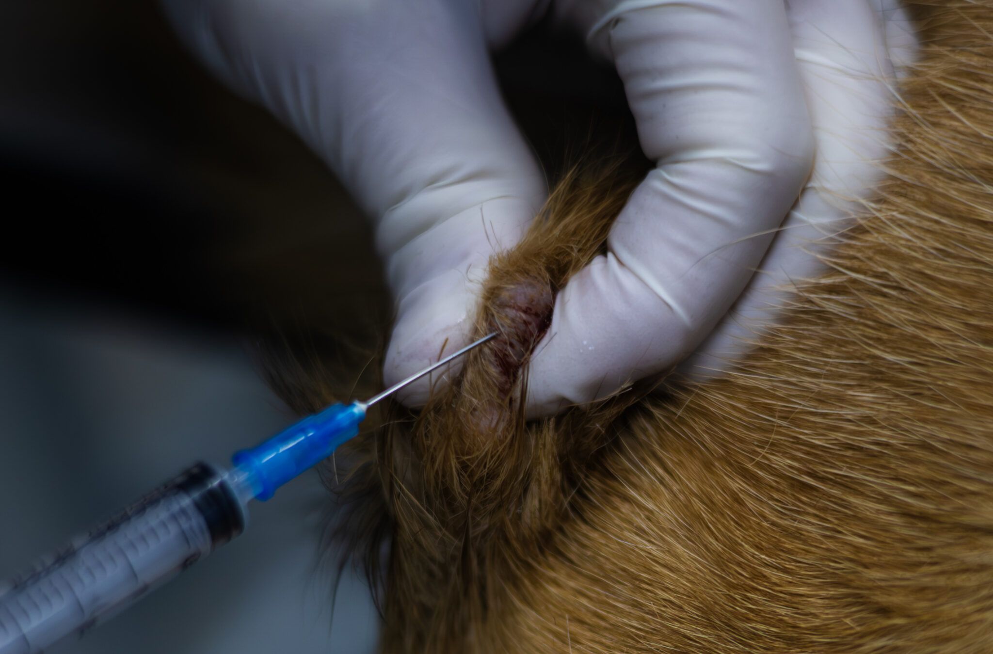
A veterinarian cannot confirm which growth your dog has by appearance alone. A biopsy remains the only reliable diagnostic tool. The vet collects a tissue sample and sends it to a pathologist who identifies the cell type under a microscope. This step determines treatment direction.
If your dog develops a suspicious skin growth, schedule a vet appointment promptly. Bring photos documenting when the bump first appeared and whether it has changed in size or appearance. Your vet may recommend a fine-needle aspirate, a less invasive initial test that sometimes provides diagnostic information before committing to full biopsy.
Early veterinary evaluation changes outcomes. Dogs with MCTs caught in early stages have better prognoses