Skin bumps on dogs spark worry in many pet owners, but not all growths signal cancer. Two conditions look nearly identical to untrained eyes yet differ dramatically in their nature and prognosis: mast cell tumors (MCTs) and histiocytomas.
Mast cell tumors are cancerous growths that develop from mast cells, immune cells in the skin. These tumors appear as raised, often itchy bumps that can range from small to large. They carry real health risks and require veterinary treatment. Histiocytomas, by contrast, are benign skin tumors derived from histiocytes, another type of immune cell. These growths are not cancerous and often resolve on their own.
The visual similarity between these two conditions creates diagnostic challenges for owners. Both appear as rounded bumps on the skin, sometimes with redness or inflammation. A dog might have a growth that looks suspicious, but only a veterinary examination with tissue analysis can definitively determine which condition is present.
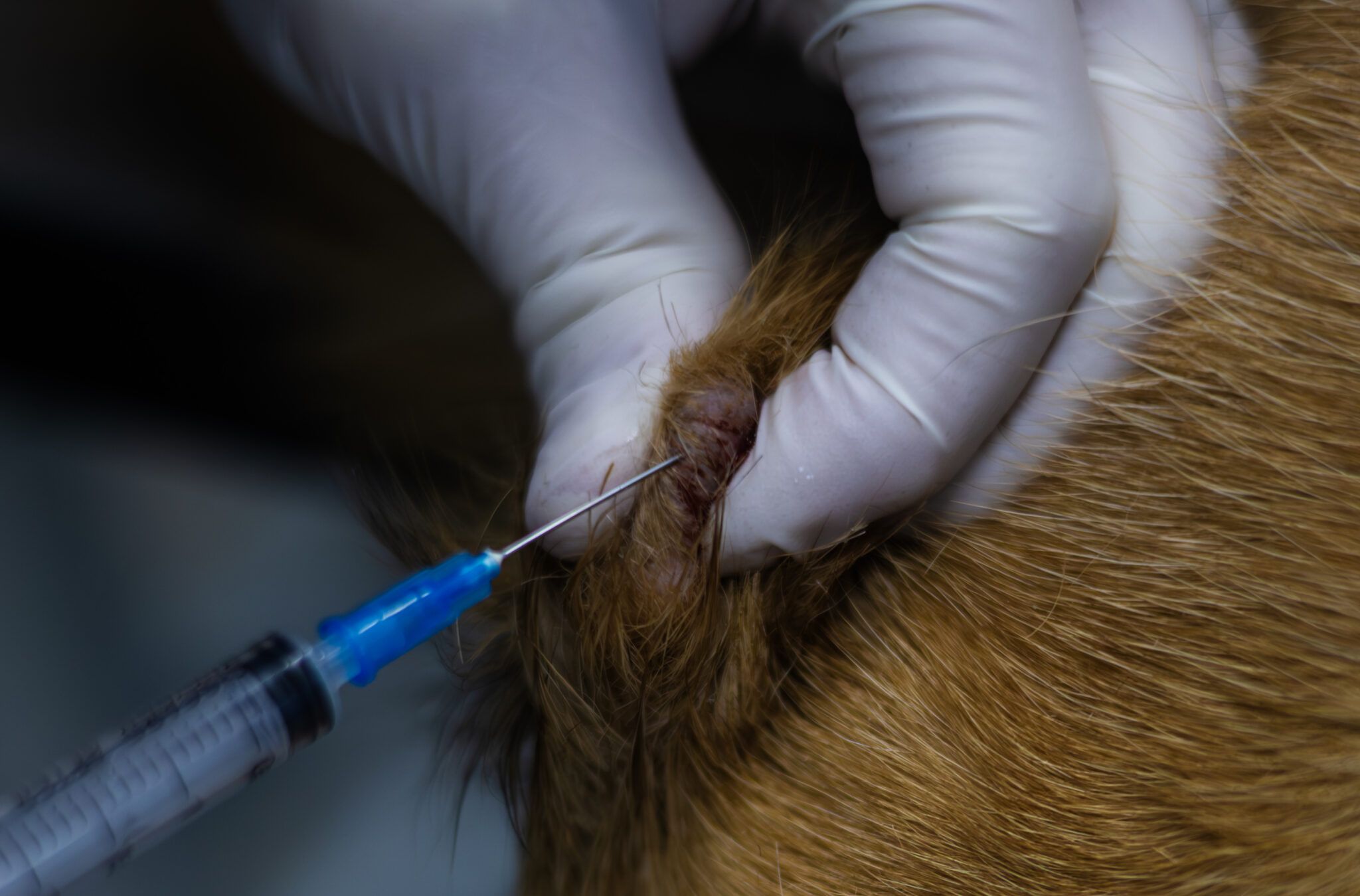
Your veterinarian will likely recommend a fine-needle aspirate or biopsy to distinguish between them. A fine-needle aspirate involves collecting cells with a small needle and examining them under a microscope. A biopsy removes a tissue sample for more detailed analysis. These procedures provide certainty about what you're dealing with.
If your dog develops any new skin growth, schedule a veterinary appointment promptly. While histiocytomas don't typically require treatment beyond monitoring, mast cell tumors demand action. Treatment options for MCTs include surgical removal, radiation, chemotherapy, or targeted medications depending on the tumor's grade and stage.
Early detection matters. Catching an MCT in its early stages generally improves outcomes. Your vet can monitor existing growths during regular check-ups and recommend treatment if changes occur.
Don't let concern spiral into anxiety if your dog