Skin bumps on dogs range from harmless to serious, and two growths in particular can confuse even careful owners. Mast cell tumors (MCTs) are cancerous, while histiocytomas are benign. The problem: they look nearly identical to the naked eye.
Mast cell tumors develop from mast cells, immune system cells that live in the skin. These tumors are malignant and can spread to other parts of a dog's body. They occur most often in middle-aged and older dogs. Certain breeds like Boxers, Bulldogs, and Labrador Retrievers face higher risk.
Histiocytomas, by contrast, originate from histiocytes, a different type of immune cell. These benign growths typically appear in younger dogs under three years old. Unlike MCTs, histiocytomas rarely spread and often shrink or disappear on their own over time.
Both tumors present as firm, raised bumps on the skin. They may be red or pink and can develop rapidly. This visual similarity creates real uncertainty for dog owners who notice a new growth. A dog could have either condition, and without a veterinary diagnosis, there is no way to tell the difference based on appearance alone.
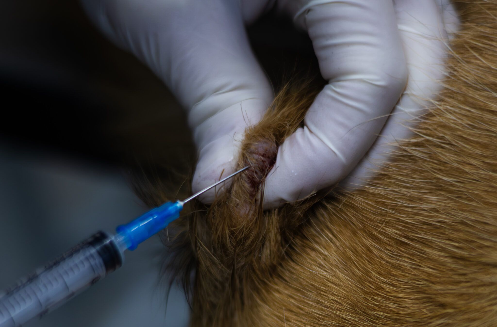
Your veterinarian can distinguish between them using a fine needle aspiration (FNA) or biopsy. An FNA involves inserting a thin needle into the growth to collect cells for microscopic examination. A biopsy removes a tissue sample for lab analysis. Either test confirms whether the growth is cancerous.
If your dog develops a skin bump, contact your veterinarian promptly. Early diagnosis matters for mast cell tumors, since treatment options like surgical removal or chemotherapy work best when caught early. Knowing whether your dog has an MCT or histiocytoma allows your vet to recommend